For US Audiences Only
FOR U.S. HEALTHCARE PROFESSIONALS
For US Audiences Only
ABOUT CYSTINOSIS

Cystinosis is an autosomal recessive lysosomal storage disease caused by mutations in the CTNS gene.1

CTNS encodes a cystine-specific transporter, cystinosin, which normally transports the amino acid cystine out of lysosomes.2,3

CTNS mutations result in absent or defective cystinosin, which prevents the normal transport of cystine out of the lysosomes. This, in turn, results in cystine crystal accumulation within lysosomes.1,3

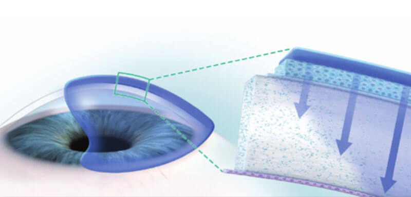
Cystine crystal formation negatively impacts many organs throughout the body and leads to significant health complications. One of the organs impacted is the eye.4,5 In the eye, cystine crystals appear most often in the cornea and conjunctiva.9

Cystinosis ocular symptoms
LOOKING AT CORNEAL CYSTINE CRYSTALS

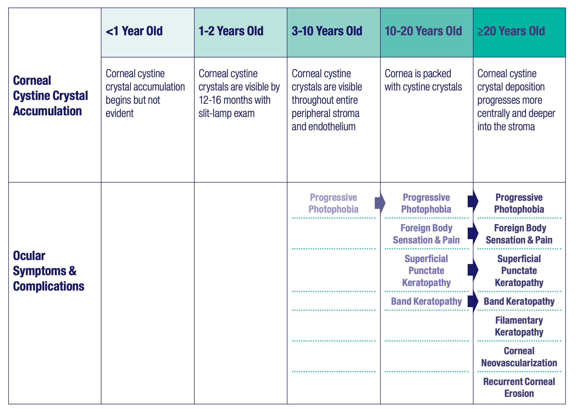
Although ocular symptoms may appear later in life than many other symptoms of cystinosis, ocular symptoms can impair quality of life in untreated patients, especially as cystine crystals accumulate over time.5,6,10

Cystine crystal deposition begins in the peripheral cornea, and then progresses more centrally and more deeply into the stroma with age.8 Corneal crystals typically appear as pointed and needle-shaped, resembling shards of glass.7,9
 

CORNEAL CYSTINE CRYSTALS
Images produced using in vivo confocal microscopy (IVCM)11

Cystinosis ocular symptoms

*Reproduced by permission from Labbe 2014

Corneal cystine crystals are not detectable at birth, but they can generally be observed via slit-lamp examination by the age of 12-16 months.12,14 As the disease progresses, by approximately 7 years of age, the entire peripheral stroma and endothelium accumulate crystals. By approximately 20 years of age, crystals can be seen in the entire corneal stroma.14


CORNEAL CYSTINE CRYSTAL ACCUMULATION AND SYMPTOMS BY AGE GROUP
(WITHOUT TREATMENT) 5,6,10,13,14,15,16

Cystinosis ocular symptoms

TARGETED TREATMENT FOR CORNEAL CYSTINE CRYSTALS IS NEEDED.

Cysteamine eye drops may effectively deplete corneal cystine crystals and, as a result, decrease or lessen the risk of developing uncomfortable or debilitating symptoms.8,14,18 Due to the cornea’s avascularity, systemic cysteamine does not affect corneal cystine crystal accumulation.12,18,19

MEASURING CORNEAL CYSTINE CRYSTALS USING IN VIVO CONFOCAL MICROSCOPY (IVCM)6,11
  • IVCM provides high resolution images of cystine crystals by corneal layer at the cellular level.
  • An IVCM score quantifies cystine crystal deposition in each layer of the central cornea, the sum of which results in the IVCM total score.
  • Higher scores designate larger amounts of cystine crystal deposits; a decrease in IVCM total score indicates a reduction in corneal cystine crystals.
IN VIVO CONFOCAL MICROSCOPY (IVCM)6

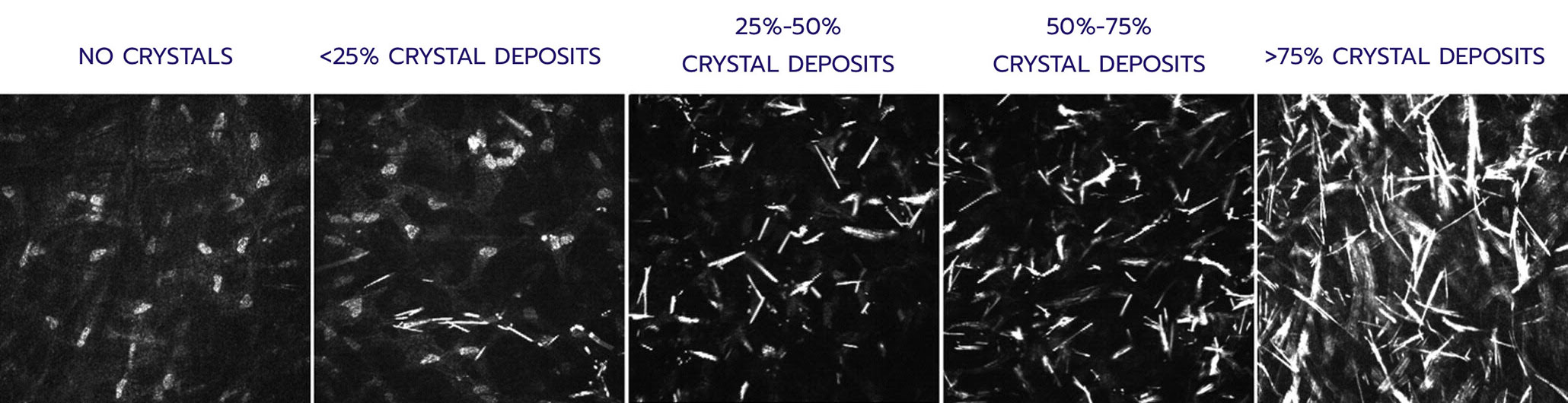
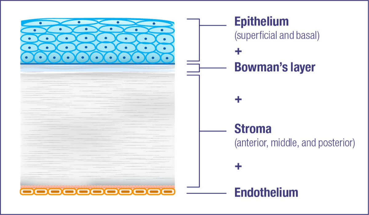
Quantifies: Corneal cystine crystal density at the cellular level in 7 layers of the cornea

IVCM Scoring: 0-4 in each layer

  • 0 = no crystals
  • 1 = <25% deposits in images
  • 2 = 25% to 50% deposits in images
  • 3 = 50% to 75% deposits in images
  • 4 = 75% to 100% deposits in images

Total score: Sum of each layer’s mean score (0-28)

Layers of the cornea
X

INDICATIONS AND IMPORTANT SAFETY INFORMATION

Important Safety Information

  • To minimize the risk of contamination, do not touch the dropper tip to any surface. Keep bottle tightly closed when not in use.

References

  1. Besouw MTP, Levtchenko EN. Improving the prognosis of nephropathic cystinosis. Int J Nephrol Renovasc Dis.2014;7:297-302. doi: 10.2147/IJNRD.S37603
  2. Goodyer P. The history of cystinosis: lessons for clinical management. Int J Nephrol. 2011;2011:1-6. doi: 10.4061/2011/929456
  3. Gahl WA, Balog JZ, Kleta R. Nephropathic cystinosis in adults: natural history and effects of oral cysteamine therapy. Ann Intern Med. 2007;147:242-250. doi: 10.7326/0003-4819-147-4-200708210-00006
  4. Baumner S, Weber LT. Nephropathic cystinosis: symptoms, treatment, and perspectives of a systemic disease. Front Pediatr. 2018; 6:1-8. doi: 10.3389/fped.2018.00058
  5. Nesterova G, Gahl WA. Cystinosis. 2001 Mar 22 [Updated 2017 Dec 7]. In: Adam MP, Ardinger HH, Pagon RA, et al., editors. GeneReviews® [Internet]. Seattle (WA): University of Washington, Seattle; 1993-2020. Available from: https://www.ncbi.nlm.nih.gov/books/NBK1400/
  6. Liang H, Baudouin C, Hassani RTJ, Brignole-Baudouin F, Labbé A. Photophobia and corneal crystal density in nephropathic cystinosis: an in vivo confocal microscopy and anterior-segment optical coherence tomography study. Invest Ophthalmol Vis Sci. 2015:56(5):3218-3225. doi: 10.1167/iovs.15-16499
  7. Gahl WA, Thoene JG, Schneider JA. Cystinosis. N Engl J Med. 2002;347(2):111-121. doi: 10.1056/NEJMra020552
  8. Shams F, Livingstone I, Oladiwura D, Ramaesh K. Treatment of corneal cystine crystal accumulation in patients with cystinosis. Clin Ophthalmol. 2014;8:2077-2084. doi: 10.2147/OPTH.S36626
  9. Emma F, Nesterova G, Langman C, Labbé A, Cherqui S, Goodyer P, Janssen MC, Greco M, Topaloglu R, Elenberg E, Dohil R, Trauner D, Antignac C, Cochat P, Kaskel F, Servais A, Wuhl E, Niaudet P, Van’t Hoff W, Gahl W, Levtchenko E. Nephropathic cystinosis: an international consensus document. Nephrol Dial Transplant. 2014;29(Suppl 4):87-94. doi: 10.1093/ndt/gfu090
  10. Tsilou ET, Rubin BI, Reed GF, Iwata F, Gahl W, Kaiser-Kupfer MI. Age-related prevalence of anterior segment complications in patients with infantile nephropathic cystinosis. Cornea. 2002;21:173-176. doi: 10.1097/00003226-200203000-00009
  11. Labbé A, Baudouin C, Deschenes G, Loirat C, Charbit M, Guest G, Niaudet P. A new gel formulation of topical cysteamine for the treatment of corneal cystine crystals in cystinosis: the Cystadrops OCT-1 study. Mol Genet Metab. 2014;111(3):314-320. doi: 10.1016/j.ymgme.2013.12.298
  12. Wilmer MJ, Schoeber JP, van den Heuvel LP, Levtchenko EN. Cystinosis: practical tools for diagnosis and treatment. Pediatr Nephrol. 2011;26(2):205-215. doi: 10.1007/s00467-010-1627-6
  13. Elmonem MA, Veys KR, Soliman NA, van Dyck M, van den Heuvel LP, Levtchenko E. Cystinosis: a review. Orphanet J Rare Dis. 2016; 11:47. doi: 10.1186/s13023-016-0426-y
  14. Tsilou E, Zhou M, Gahl W, Sieving PC, Chan C-C. Ophthalmic manifestations and histopathology of infantile nephropathic cystinosis: report of a case and review of the literature. Surv Ophthalmol. 2007;52(1): 97-105. doi: 10.1016/j.survophthal.2006.10.006
  15. Gahl WA, Kuehl EM, Iwata F, Lindblad A, Kaiser-Kupfer MI. Corneal crystals in nephropathic cystinosis: natural history and treatment with cysteamine eyedrops. Mol Genet Metab. 2000;71(1-2):100-120. doi:10.1006/mgme.2000.3062
  16. Pinxten A-M, Hua M-T, Simpson J, Hohenfellner K, Levtchenko E, Casteels I. Clinical practice: a proposed standardized ophthalmological assessment for patients with cystinosis. Ophthalmol Ther. 2017;6:93-104. doi: 10.1007/s40123-017-0089-3
  17. Hala A Helmi, Jeylan El Mansoury, Selwa Al Hazzaa, Abdulaziz Al Zoba, Qais S Dirar. Asymmetrical Ocular Manifestations of Nephropathic Cystinosis; A Case Report: Am J Case Reports. 2019 Sep 4;20:1308-1313. doi: 10.12659/AJCR.916737
  18. Nesterova G, Gahl W. Nephropathic cystinosis: late complications of a multisystemic disease. Pediatr Nephrol. 2008;23: 863-878. doi: 10.1007/s00467-007-0650-8
  19. Willoughby CE, Ponzin D, Ferrari S, Lobo A, Landau K, Omidi Y. Anatomy and physiology of the human eye: effects of mucopolysaccharidoses disease on structure a function – a review. Clin Exp Ophthalmol. 2010;38:2-11. doi: 10.1111/j.1442-9071.2010.02363.x