About CYSTADROPS
LEARN MORE
Cystinosis is a rare genetic disorder caused by mutations in the CTNS gene.
The job of the CTNS gene is to make cystinosin. Cystinosin transports, or carries, the amino acid cystine out of the lysosomesLysosomes are the organelles within the cells in your body that act as cell digestive system. Lysosomes break apart amino acids such as cystine.
In those with cystinosis, the cystinosin transport process does not work correctly. As a result, cystine builds up inside lysosomes. Over time, the cystine forms into crystals.
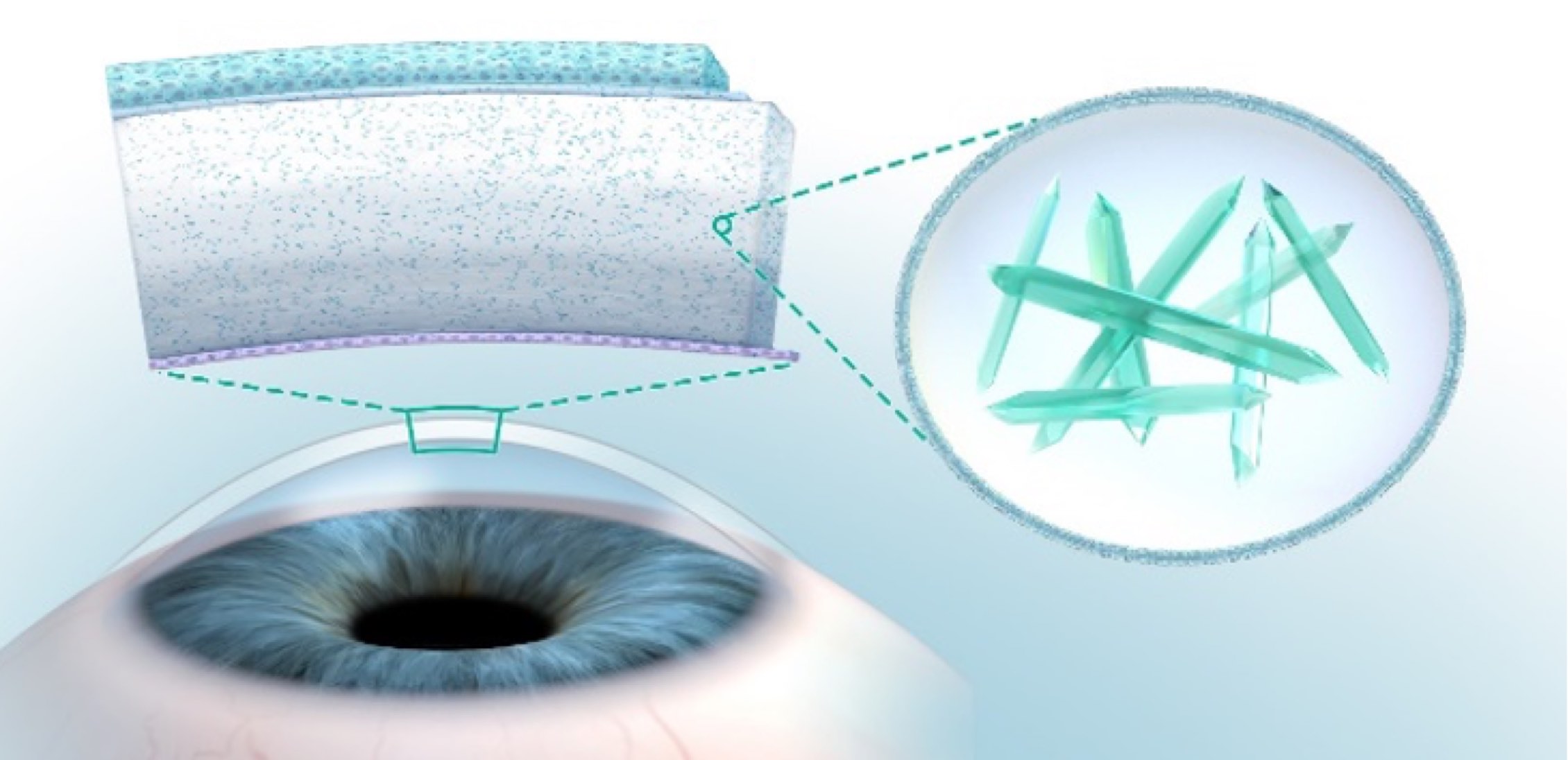
Cystine crystals may build up in cells in every organ of your body, including your eyes. The corneathe front layer of the eye is the part of the eye that may be affected the most.
In the cornea, the cystine crystals are usually pointed and needle-shaped and look like shards of glass.
The corneathe front layer of the eye has very important functions:
The cornea has several layers, and cystine crystals can build up throughout these layers. Without proper treatment, cystine crystals will continue to form and will progress deeper into all layers of the cornea.
At first, as cystine crystals begin to build up in your corneathe front layer of the eye, you may not notice any eye symptoms. But gradually, crystal build-up progresses deeper into your cornea, affecting all the layers, and you may begin to feel some discomfort.
Symptoms may include:
Photophobia and other symptoms can be uncomfortable and even painful. These symptoms can also affect your ability to carry out daily activities.
Some people use a small notebook to keep track of symptoms over time. This can be helpful when you talk to your doctor.
Talk to your doctor about your or your child’s eye symptoms and how these symptoms affect daily activities.

To measure the amount of cystine crystals in your corneathe front layer of the eye, your eye doctorAn ophthalmologist or optometrist. can use a machine called a slit-lamp microscopeA slit lamp is a microscope with a bright light used during an eye exam. It gives your eye doctor a closer look at the different tissues at the front of your eye and inside your eye.. This microscope directs a high-intensity beam of light onto your cornea, which allows your doctor to score the amount of cystine crystals that have built up and to monitor changes over time. Your eye doctor can also use a different imaging machine to evaluate the corneal cystine crystals.
The slit-lamp microscope can also be used to assess photophobia. Your doctor may also ask questions about how photophobia may be affecting your daily life.
It’s important to know that cystine-depleting oral medicinesmedicines taken by mouth do not reduce the build-up of cystine crystals in your corneathe front layer of the eye. These medicines are delivered to organs in your body through your bloodstream. But these medicines cannot reach your cornea since it does not have blood vessels.
Without proper treatment, more and more crystals will form in your cornea and progress deeper into the layers of the cornea.
Daily treatment with cysteamine eye drops is the only way to deplete the build-up of corneal cystine crystals and reduce eye symptoms caused by cystinosis.
CYSTADROPS (cysteamine ophthalmic solution) 0.37% is a cystine-depleting agent indicated for the treatment of corneal cystine crystal deposits in adults and children with cystinosis.
INDICATIONS AND IMPORTANT SAFETY INFORMATION